
Need for Transport
- carry required substances to body parts
- remove metabolic wastes from the body
- Lower organisms and simple multicellular organisms have small bodies that make them have a large surface area to volume ratio. Diffusion alone is enough to transport substances.
- Higher organisms have large bodies and thus smaller surface area to volume ratio. Hence, tissues and organs are far from the site of supply of materials. Therefore, they require an elaborate transport system.
TRANSPORT IN PLANTS
- Simple plants such as mosses and liverworts transport substances by diffusion, osmosis, and active transport.
- Higher plants have a specialized transport system known as the vascular bundle. It comprises of xylem (transports water and mineral salts) and phloem (transports dissolved food substances).
Root Structure and Function
The primary functions of roots are anchorage and absorption. Specialized roots store food and water, and allow gaseous exchange.
- Root cap: protects the apical meristem.
- Root hairs: microscopic outgrowths of epidermal cells. Are thin walled to shorten distance for absorption of water and mineral salts. Have an elongated cytoplasm at one side to increase surface area for absorption of water and mineral salts.
- Piliferous layer: a special epidermis of young roots that gives rise to root hairs.
- Cortex: loosely packed thin walled cells to allow passage of water molecules to reach the vascular bundles; and to store substances.
- Endodermis: a single layer of cells surrounding the vascular bundles, characterized by possession of starch grains and a casparian strip, it controls the amount of water and mineral salts entering into the vascular bundles.
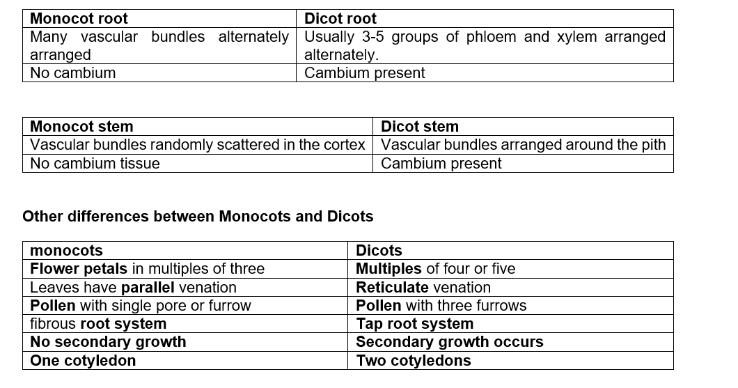
- Vascular bundles: each vascular bundle consists of xylem and phloem. In monocotyledonae, xylem alternate in the arrangement with phloem. In Dicotyledonae, the xylem is star-shaped and the phloem is located at the centre of the star.
- Epidermis: a single layer of cells covering all other inner tissues
Stem Structure and Function
The primary functions of stems are support, conduction of water and mineral salts and manufactured food. Other functions of specialized stems include storage of food and water, gaseous exchange and perennation.
- Epidermis
- Cortex: simple tissues found in the cortex are:
Collenchyma: walls thickened at the corners with cellulose and pectin deposits, and thus it is a strengthening tissue;
Parenchyma: irregular cells, thin walled and loosely packed, with intercellular air spaces, store water and food. Some parenchyma cells contain chloroplasts and are called
Sclerenchyma: walls thickened by lignin, serve as a strengthening tissue.
3. Pith: central region with parenchyma cells that store water and food substances.
4. Vascular bundles
Comparison between the Structure of Monocots and Dicots
Absorption of Water and Mineral Salts
Absorption of Water
- Water is drawn into root hair cells by osmosis
- Cell sap of root hairs contains dissolved substances that make its concentration to be greater than that of the soil solution
- Therefore, there exists a concentration gradient between cell sap and soil water
- This exerts a higher osmotic pressure thus drawing water molecules from soil across the cell wall and cell membrane of root hair cells
- The osmotic forces exerted by the cells overcome the water retaining powers of the soil thus water enters the root hair cells
- As more water is drawn into root hair cells, it dilutes the cell sap making it less concentrated than that in the adjacent cortex cell of the root
- Due to osmotic gradient, water moves from the adjacent cells to the next by osmosis
- Similarly, water passes through the successive cortex cells until it enters the xylem vessels located in the center of the root
- The xylem vessels then conduct the water up into the xylem vessels of the stem and into the leaves
Uptake of Mineral Salts
- Soil water contains dissolved mineral salts, which plants require for their growth and proper functioning
- Generally, the concentration of the cell sap in the root hairs is greater than that in the soil
- The mineral salts therefore enter the root hairs against the concentration gradient
- This process requires the use of energy and is referred to as active transport
- Active transport is believed to involve substances known as carriers
- These carriers combine with the mineral ions and then carry them across the plasma membrane into the cell
- Thus, the carriers move back and forth carrying the salt ions from the soil water to the root hair cells
- After absorption, the mineral salts move through the root cells into the xylem vessels of the vascular tissue in the centre of the root
- Then the salts and water are carried up the stem into the leaves by a combination of osmosis, diffusion, root pressure, transpiration pull, cohesive forces and capillary attraction
Forces involved in Transportation of Water and Mineral Salts
- Transpiration pull
- A suction force pulls a stream of water from the xylem vessels in the stem and roots. It develops when water vaporizes from the spongy mesophyll cells, increasing the concentration of their cell sap, which in turn increases the osmotic pressure and causes water to flow from surrounding cells and xylem vessels.
- Transpiration pull creates a continuous stream of water flowing from the roots, up the stem to the evaporating surface referred to as transpiration stream
Distinguish between the terms transpiration pull and transpiration stream
[1]How does transpiration pull arise? [3]
- Cohesion and Adhesion forces
- Cohesion force: forces that keep water molecules together
- Adhesive force: attractive force between water molecules and the walls of xylem vessels
- Capillarity
- Ability of a solution to rise up in a tube with a very small diameter because of surface tension
- Water rises up by capillary action in xylem vessels because they are very narrow and there is a high attractive force between the water molecules and the cell walls
- Root pressure
- A force in the roots that pushes water up the stem
- Root pressure is caused by active pumping of water across the endodermis to the xylem vessels because the casparian strip present in endodermis is almost impermeable to water
- Energy is required in this process
TRANSPIRATION
Dfn: Process by which plants lose water in the form of water vapour into the atmosphere
- Sites of transpiration are stomata (80-90%), cuticle (up to 20%), and lenticels (negligible).
- Each stoma opens into the sub-stomatal air spaces that are lined with spongy mesophyll cells.
- Water vaporizes from the spongy mesophyll cells into the sub-stomatal air spaces, and then the water vapour diffuses out of the leaf through the stomata
- The cell sap of the spongy mesophyll cells becomes more concentrated than the adjacent cells
- Therefore, this increases the osmotic pressure of the cells
- As a result, water flows into the cell from other surrounding cells, which in turn take in water from xylem vessels within the leaf veins
Factors that Increase Transpiration Rate
- Thick layer of cuticle: increases diffusion distance
- Broad leaves: expose a large surface area
- More stomata on upper leaf surface/ large stomatal apertures: exposed to sun light, wind and heat
- High temperature: increases capacity of atmospheric air to hold more water vapour; increases internal temperature of leaf, which in turn increases LHV and therefore enhances evaporation from the leaf cells.
- Low relative humidity: saturation deficit is high; more water vapour diffuses out of the leaf into the atmosphere
- Wind: carries away water vapour around leaves, maintaining a high diffusion gradient between the inside and outside of the leaf
- High light intensity: causes stomata to open fully, exposing sub-stomatal air into direct contact with the outside environment.
- Low atmospheric pressure/high intercellular air pressure: more water vapour is lost to the atmosphere due to the pressure deficit in the atmosphere
- Adequate water supply from the soil: walls of mesophyll cells are kept continuously wet
Factors that Decrease Transpiration Rate
- Thin cuticle
- Small leaves/ needle-like
- Few stomata on upper leaf surface/sunken stomata
- Midday closure: protects from wilting
- Reversed stomatal rhythm: to conserve water in xerophytes
- Leaf fall: reduced S/A
- Hairy leaves/scales on leaves: trap still moist air.
- Low temperature
- High relative humidity
- Still air
- Low light intensity
- High atmospheric pressure/low intercellular air pressure
- Lack of enough water in soil
Guttation
- It is the loss of water in liquid form through hydathodes
- common in hydrophytes when the relative humidity is high
- caused by root pressure
- typically occurs during the night
- occurs only at the tip or along the edges of leaf
- The soil must have high amount of water than that in the roots
- When the water evaporates, sugars and minerals left behind
Significance of Transpiration
- Replaces lost water
- transports water and mineral salts
- cools plants
- removes excess water in aquatic plants
- maintains turgor
Structure and Function of Xylem Tissue
- Xylem tissue is made up two main cell types: vessels and
- Xylem vessels
- Long hollow tubes to allow flow of water and dissolved mineral salts.
- Non-living
- Walls strengthened by deposition of lignin to prevent collapsing.
- Have bordered pits along their walls to permit passage of water in and out of the lumen into the neighboring cells.
- Found in angiosperms only (flowering plants)
2. Tracheids
- Hollow cells
- Non-living
- Have lignin deposits on their walls.
- Have tapering / chisel-shaped ends
- Sloping end walls are perforated.
- Have pits on the sidewalls to allow lateral water movement to cells surrounding the xylem. This makes them less efficient than vessels in conducting water than the vessels.
- Tracheids perform two functions: support and transport of water.
- Found in Pteridophytes, gymnosperms and angiosperms
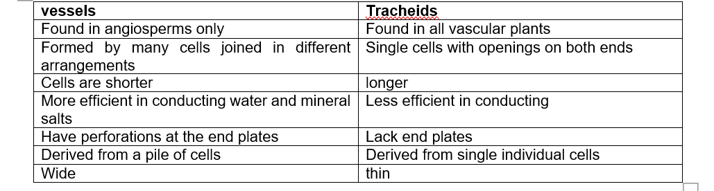
Comparison between Vessels and Tracheids
Translocation of Organic Compounds
- Translocation is the transport of soluble organic products of photosynthesis within the plant.
- Occurs in phloem tissue
Phloem structure and Function
- Cytoplasmic filaments are contractile in nature to push food material from one sieve tube to another
- Sieve tubes are hollow to allow passage of food material
- Companion cells are densely packed with mitochondria, which produce energy for active transport of food
- Presence of sieve pores allows the passage of food from one sieve tube to the next.
- The plasmodesmata connect sieve cells with companion cells and allow for exchange of materials between the two cells
Q Describe the function of the endodermis in roots [3]
Q State two ways in which the structure of a phloem sieve tube is adapted for the transport of assimilates [2]
TRANSPORT IN ANIMALS
- Open circulatory system: transport fluid (haemocoel) is contained in the general body cavity or coelom, ex arthropods
- Closed circulatory system: transport fluid (blood) is conveyed in blood vessels, ex vertebrates, annelids.
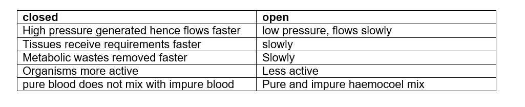
Comparison between Open and Closed Circulatory System
Q Explain why the mammalian circulatory system is described as a closed double circulation.
Transport in a typical Insect (cockroach)
- Has open circulatory system where blood is contained in coelom and within a dorsal tubular heart.
- Blood contains suspended leucocytes and some pigments.
- Transport of gases occurs mainly by diffusion in the tracheal system.
- The heart has thirteen chambers, three in the thorax and ten in the abdominal segments.
- The anterior segment is joined to the aorta that empties the blood into sinuses of the head.
- Each chamber contains a pair of valves at the anterior part, which prevent back flow of the blood.
- Each chamber has a pair of lateral openings called ostia, which are closed by valves. The valves allow blood to flow into the heart through the ostia but not out of it.
The Mammalian Circulatory System
- organization of the transport system in mammals: Heart→arteries→arterioles→capillaries (in tissues)→venules→veins→heart
- Mammals have a double circulation: blood flows into the heart twice for every complete circulation. First, blood is pumped to the lungs for oxygenation and then back to the heart (pulmonary circulation). Then blood is pumped from the heart to the rest of the body organs (systemic circulation).
- Some animals like fish have single circulation in which blood flows only once through the heart for every complete circuit.
The Structure and Function of the Heart
- Pericardium: membrane that encloses heart to prevent over stretching, secretes pericardial fluid to lubricate the heart wall when pumping blood
- Cardiac muscles have interconnected fibres so that waves of contraction can travel throughout the muscle
- Cardiac muscles have myogenic property to initiate contraction without external nerve supply; and to contract continuously without fatigue
- Sino-atrial node on the right atrium initiates contractions/serves as the pacemaker
- Purkinje fibres allow waves of contraction to reach ventricles
- Coronary artery supplies the heart muscle with nutrients and oxygen
- Coronary vein removes metabolic wastes
- Right auricle: thin walled, receives deoxygenated blood from through vena cavae veins
- Left auricle: thin walled, receives oxygenated blood from the lungs through the pulmonary vein
- Left ventricle: has thicker muscles than right ventricle because it pumps blood to a longer distance to all the body parts which requires higher pressure
- Right ventricle has thinner muscles because it pumps blood a shorter distance from heart to the lungs which requires less pressure
- Inter-ventricular septum prevents mixing of oxygenated and deoxygenated blood
- Atrio-ventricular valves prevent blood from flowing back into the auricles when ventricles contract. (left: bicuspid valves; right: tricuspid valves)
- Tendons prevent valves from turning inside out when ventricles contract
- Semi-lunar valves prevent back flow of blood when the ventricles relax
Q Explain how increased intensity of exercise leads to an increased heart rate. [4]
Pumping Mechanism of the Heart
Diastole (Relaxation)
- Ventricle muscles relax
- Volume of ventricle increases pressure decreases
- Cuspid valves open
- Deoxygenated blood from body tissues flows into the right ventricle
- Oxygenated blood from lungs flows from the left atrium into the left ventricle
- Semi-lunar valves close preventing blood from flowing back into the ventricles
- The slight contractions of the auricles force the blood into the ventricles
Systole (Contraction)
- Ventricle muscles contract
- Volume of ventricles decreases while pressure increases
- Blood is forced out of the heart to the lungs and to body tissues
- Cuspid valves close preventing blood from flowing back into the auricles
Initiation of Heartbeat
- Heartbeat is initiated by sinoatrial node (SAN) or the pacemaker found at the right atrium
- SAN starts electrical waves that spread over the walls of the two atria making them contract
- waves then pass through atrioventricular node (AVN)
- There is a short delay of about 0.1 seconds before waves pass from atria to ventricles, to prevent them from contracting simultaneously (it would be disastrous if this happened)
- AVN passes waves on to Purkinje tissue in the interventricular septum
- to the apex of the heart
- and then through the ventricle walls
- causing ventricles to contract from the base upwards
- forcing blood to flow out thro the vessels leaving the heart
- Vagus nerve slows down heartbeat while sympathetic nerve speeds it up
Q Describe the initiation and control of heartbeat (8 marks)
Structure and Function of Blood vessels
- Arteries
- Thick muscular wall to resist pressure
- smooth endothelium to reduce friction as blood flows
- Muscular layer to produce pulsating action to force blood forwards as heart beats
- Muscular wall controlled by nerves and hormones to alter lumen diameter so as to regulate blood flow to organs
- Capillaries
- Numerous and very close to tissues to increase S/A for exchange of substances
- One cell-thick wall to reduce diffusion distance
- Very narrow lumen to bring substances very close to cells/tissues for fast diffusion/create very high pressure for ultra filtration
- Smooth endothelium to reduce friction to blood flow
- Veins
- Larger lumen than arteries to offer minimum resistance to blood flow
- Valves to prevent back flow of blood (skeletal muscular contraction assists in blood flow)
Diseases and Defects of the Circulatory System
- Thrombosis: formation of blood clot in vessels, commonly due to heavy fat intake e.g. coronary thrombosis
- Arteriosclerosis: deposition of calcium in walls of blood vessels leading to hardening, associated with being overweight, lack of exercise and emotional stress
- Varicose veins: swollen superficial veins at the back of legs due to failure of valves to function properly; results in the retention of tissue fluid
- Hypertension or High Blood Pressure: pressure above upper limit of normal blood pressure (140/90)
- associated with intake of too much salt in food, sustained stress and arteriosclerosis
Structure and Functions of Mammalian Blood
- consists of fluid called plasma and cells suspended in it
- plasma consists of dissolved substances and plasma proteins
- Serum is blood plasma without fibrinogen and cells
- Functions of plasma: Transport cells; dissolved food substances; metabolic wastes; hormones; some oxygen and CO2; regulate pH of body fluids; distribute heat
Red blood cells
- No nucleus when mature
- Short life span
- Made in bone marrow of short bones in adults, liver and spleen in the embryo
- Contain haemoglobin (Hb)
- Cytoplasm red/pink
- Number increases when living at higher altitudes
- Main function is transport
Adaptations
- no nucleus when mature: create more space to pack more haemoglobin to carry more oxygen (unfortunately makes RBCs have short life span)
- haemoglobin to transport oxygen and CO2
- numerous in number to increase S/A for transport
- flexible plasma membrane to enable them squeeze through narrow capillaries
- biconcave shape increases S/A over which gaseous exchange can take place
- carbonic anhydrase speeds up the conversion of CO2 to weak carbonic acid
Q Mature mammalian red blood cells have no nuclei. State one advantage and one disadvantage of this [2]
White Blood Cells (leucocytes)
- nucleated
- very long life span
- lack Hb
- cytoplasm colorless
- fewer in number than RBCs
- formed in bone marrow of long bones and in lymph nodes
- their number increases during infection
- main function is defense
Q State five differences between red blood cells and white blood cells (5 marks)
Defense Function of Leucocytes
- Phagocytosis
- Antibody secretion: antitoxins neutralise toxins; agglutinins clump together microorganisms; lysins digest cell walls and membranes of microorganisms; opsonins coat microorganisms making them easier to phagocytose
Q State four ways antibodies function to fight pathogens [4]
Platelets (Thrombocytes)
- Fragments from large cells in the bone marrow
- No nucleus
- Take part in blood clotting
Transport of Oxygen and Carbon (IV) oxide
Oxygen
- Haemoglobin in RBCs has high affinity for oxygen
- In the lungs, there is high oxygen tension
- haemoglobin picks up oxygen to form an unstable cpd called oxyhaemoglobin
- Oxyhaemoglobin is transported to tissues where there is low oxygen tension
- In tissues, oxyhaemoglobin readily dissociates into haemoglobin and oxygen
- Oxygen diffuses out of RBCs and through the capillary walls into the tissues
- Free haemoglobin is transported to lungs to pick up more oxygen molecules
- Foetal Hb has higher affinity for oxygen than maternal Hb
Carbon (IV) oxide
- RBCs transport about 95% of CO2
- carbonic anhydrase inside speeds up the reaction btn CO2 and water to form weak carbonic acid
- Carbonic acid dissociates into hydrogen carbonate and hydrogen ions
- hydrogen carbonate ions diffuse out of RBCs into plasma and is transported to the lungs
- to counterbalance charge, chloride ions diffuse into the RBCs cytoplasm (chloride shift)
- Hydrogen ions are buffered by i) Hb forming weak haemoglobinic acid, and ii) plasma proteins forming proteinic acids
- Some CO2 that diffuses into RBCs combines with the amino acids of Hb to form carbaminohaemoglobin cpd
- A small amount of CO2 dissolves in plasma water to form carbonic acid
- In lungs, there is low CO2 tension which favours the release of CO2 for exhalation
Q State three functions of haemoglobin [3]
Q State two advantages of transporting carbon (IV) oxide in red blood cells [2]
Q Explain the meaning of the term chloride shift [2]
Q Describe how respiratory gases are transported in the human body [12]
Q State the advantage of foetal haemoglobin having higher affinity for oxygen than maternal haemoglobin [2]
Q Red blood cells have no nucleus. Explain how this feature is an adaptation to the function of red blood cells [2]
Respiratory Poisoning by Carbon (II) Oxide (Carbon Monoxide)
- CO has higher affinity for haemoglobin than oxygen
- therefore it readily combines with Hb to form a cpd called carboxyhaemoglobin
- which being stable reduces the capacity of Hb to transport oxygen to tissues
- if a lot of CO is inhaled, the person will die of suffocation
Blood Clotting Process
- when blood vessels are injured
- the platelets exposed to air
- and rupture to release thromboplastin
- which neutralises heparin
- and activates prothrombin to thrombin
- a reaction which requires calcium ions
- formation of prothrombin requires vitamin K
- Thrombin converts fibrinogen to fibrin
- which forms a meshwork of fibers across the cut surface and dries up to form a scab
- that stops excessive bleeding
- and protects the damaged tissues from infection by pathogens
Q Describe the process that leads to the formation of a blood clot on a wound [11]
Preventing blood clotting in undamaged blood vessels
- blood contains prothrombin, but not thrombin
- blood contains heparin, an anticlotting factor
- platelets are not exposed to air
Human Blood Groups
- proteins called antigens are expressed on RBC membranes
- these are A and B and they determine type of blood gp
- some people lack A and B antigens on their RBCs and belong to blood group O
- antigens determine type of blood gp
- the plasma too has proteins called antibodies
- the antibodies do not correspond with the antigens so as to prevent agglutination /clumping of RBCs
- several other antigens expressed on RBCs have been discovered, the most important is called Rhesus factor
- people with Rhesus factor are Rh+ve while those lacking are Rh_ve
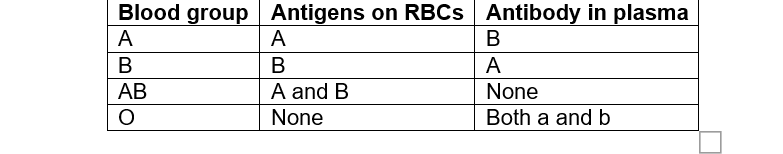
Summary of Blood Groups, Antigens and Antibodies
Q Explain why blood group B cannot be transfused to a recipient with blood group A
[4]
Suggested response
Recipient blood has antigen A and antibody b; donor blood has antigen B and antibody a; the recipient’s antibody b would react with the donor’s antigen B; this would cause agglutination of blood; blocking blood vessels which may result to death;
Blood Transfusion
- it is the transfer of blood from a donor to the circulatory system of the recipient
- gp A receives blood from gp A and gp O donors
- gp B receives blood from gp B and gp O donors
- gp AB receives blood from gp AB, gp A, gp B, and group O donors
- blood gp AB is referred to as a universal recipient while blood gp O is called universal donor
- donated blood is mixed with an anticoagulant and kept at very low temperature
- the blood is not used after one month because most of the RBCs will have died
- the blood gp of donor and recipient are determined to ensure they are compatible, this is called blood group typing
- donor’s blood is tested for pathogens such as HIV, Hepatitis B and this is called blood screening
- cross matching is done to check if minor blood groups can cause serious agglutination of recipient’s blood
Rhesus incompatibility and its significance in Pregnancy
- if a Rh- mother bears a Rh+ baby, fragments of foetal RBCs can pass across the placenta into mother’s blood stream
- mother’s immune system responds by producing rhesus antibodies
- that in turn pass across the placenta into the blood of foetus
- but the antibodies formed are too few to affect the first pregnancy
- therefore the first baby is born without any serious reaction
- but during subsequent pregnancies bearing Rh+ foetus, there occurs a severe antigen-antibody rxn
- between rhesus antigens on foetal RBCs and maternal rhesus antibodies
- foetal RBCs are destroyed
- causing haemolytic disease of the new-born or erythroblastosis foetalis
- the new born baby can be saved by replacing its blood with Rh- blood
- or by transfusing blood to the foetus in-utero
- the disease can be prevented by treating the mother with an anti-rhesus globulin
- which coats the surfaces of her RBCs thus preventing the antigen-antibody reactions
Q Describe the cause, treatment and prevention of haemolytic disease of the newborn [10]
The Lymphatic System
Formation of lymph
- lymph is formed by ultra-filtration of blood from blood capillaries in tissues
- the pumping force of the heart together with the narrow lumens of the capillaries exert a high pressure
- that forces the fluid part of blood to filter out of the capillary walls
- into the surrounding tissues
- This filtrate consists of all the constituents of blood plasma except the blood cells and the proteins because these are too large to filter out of capillary walls
- The fluid is known as tissue fluid or intercellular fluid
- And bathes the cells of tissues supplying them with oxygen, food and other useful substances
- cells absorb these substances and pass out CO2 and other waste products
- Most of the tissue fluid then returns to the blood system through the venule end of the capillaries
- due to the lower hydrostatic pressure of the blood at the venule end compared to that at the arteriole end of the capillary
- The excess tissue fluid drains into the lymph vessels where it is known as lymph
- Lymphatic vessels from various parts of the body join together to form thoracic duct which drains into the left subclavian vein near the neck
- Flow of lymph is aided by movement of skeletal muscles around the lymph vessels which squeeze them; inspiration movements that suck lymph into the thoracic duct; exhalation movements that force lymph into the left subclavian vein; and presence of valves that prevent back flow of lymph
- Along the lymphatic system are swellings called lymph nodes that produce lymphocytes
Q Describe the formation and transport of lymph [14]
Immune Responses
- These are reactions in the body that occur when antigens are introduced
- Immune responses involve the production of antibodies or leucocytes which combine with antigens
- An antibody is a chemical substance, usually a protein, which is formed in the blood when an antigen is introduced into the tissue of a human being or any other animal
- An antibody has a chemical composition which is complementary to the antigen against which it reacts
- Therefore, a particular antibody combines with or responds only to a particular antigen to make it harmless
- Antibodies are produced by leucocytes called lymphocytes
- When harmful organisms or proteins invade the body, some lymphocytes start producing antibodies which are complimentary to them
- The bone marrow also begins to produce more phagocytes while the thymus gland also begins to produce more lymphocytes
Types of Immunity
- Natural (innate) immunity
- inherited (transmitted from parent to offspring)
- manifested as immunity against a particular disease in a family, race or breed
- Acquired immunity
- develops after suffering from a disease or thro vaccination
- It is either natural or artificial and in both cases it can be active or passive
- active if antibodies are formed as a response to natural infection by a pathogen; and passive if antibodies are passed from mother to foetus through the placenta or to the new born through colostrum in maternal milk
- Artificial acquired immunity is either active or passive; it is active when antibodies are formed in response to vaccination; and is passive where serum containing antibodies is transferred from one animal to another, e.g. horse antiserum against snake bite confers immunity to snake bite victims
- Vaccination is the administration of dead or attenuated (weakened) pathogens or chemically treated toxins (toxoids)
- Following either a natural infection by a disease causing organism or vaccination, the antigens stimulate the immune system to produce corresponding antibodies which destroy the pathogens
- The body then retains the “memory” of the structure of specific antigens in a small number of cells called memory cells, we say the body has been immunized against the disease
- In case the same disease causing organism attacks the immunized body again these memory cells divide very rapidly into antibody secreting cells called plasma cells
- Therefore a large amount of specific antibody is produced within a relatively short time to eliminate the pathogens
NOTE:
Vaccination: refers to the act of giving a vaccine either by injection or swallowing
Immunization: refers to the process of both having the vaccine and becoming immune to the disease as a result of the vaccine
Allergic Reactions
- an allergy is a hypersensitive reaction to an antigen by the body
- it occurs when antibody-antigen combination produces a violent reaction in the body
- allergic pple are hypersensitive to usually harmless materials like dust, pollen grains, some foods, some drugs and certain air pollutants
- allergy manifests itself as skin rashes, vomiting, difficulties in breathing or sneezing
- the body reacts by over-producing antibodies against harmless antigens
- the antibody –antigen rxn takes place on the surface of mast cells that burst open
- releasing histamine
- histamine increases the permeability of epithelial cells thus making them take in fluid
- the intercellular spaces also become filled with fluid and swell up
- histamine causes inflammation and pain
- sometimes anaphylaxis occurs in which blood vessels lowering BP too much causing death
- bee stings can cause death by anaphylaxis
- avoiding the allergen or administration of antihistamine drugs can control allergic reactions
Q People may be immunised against diseases using vaccines. Which part of the vaccine stimulates the body’s defence system? [1]
Q Explain why a person has who been vaccinated against measles does not catch measles even when he comes into contact with it [3]
Q What is an allergen? [1]
Describe how histamine is produced in response to an allergen [3]