
Movement is a characteristic of all living organisms.
- Most animals move from place to place but some are sessile (i.e. fixed to the substratum).
- However, though not easily observed all living protoplasm shows movement of one type or another.
Necessity for support and movement in plants
- They enable plants to be held upright to trap maximum light for photosynthesis and gaseous exchange.
- It enables animals and plants to adjust to their environment.
- To hold flowers and fruits in appropriate position for pollination and dispersal respectively.
- To enable plants to grow to great heights and withstand forces of environment e.g. strong winds.
- Movement of male gametes to effect fertilization and ensure perpetuation of a species.
- Plant parts move in response to certain stimuli in the environment of tropisms.
Tissue distribution in Monocotyledonous and Dicotyledonous plants
- Vascular bundles are the main support tissues in plants.
- In monocotyledonous stem they are scattered all over the stem.
- while in dicotyledonous stem they are found in a ring or rings.
- In monocots the xylem and phloem alternate around with pith in the centre.
- In dicots of the xylem forms a star in the centre – there is no pith.
- Phloem is found in between the arms of xylem.
- Dicotyledonous plants have cambium which brings about secondary growth resulting in thickening of the stem and root hence providing support.
- Secondary xylem becomes wood, providing more support to the plant.
Role of support tissues in young and old plant
Plants are held upright by strengthening tissues ;
- parenchyma,
- collenchyma,
- sclerenchyma
- xylem tissue.
- Parenchyma and collenchyma are the main support tissues in young plants.
Parenchyma –
- They are found below the epidermis.
- They form the bulk of packing tissue within the plant between other tissues .
- They are tightly packed and turgid they provide support.
Collenchyma –
- Their cell walls have additional cellulose deposited in the corners.
- This provides them with extra mechanical strength.
Sclerenchyma –
- Their cells are dead due to large deposits of lignin on the primary cell wall.
- The lignified wall is thick and inner lumen is small, hence provide support.
- Sclerenchyma fibres are arranged in elongated and in longitudinal sheets giving extra support.
- They are found in mature plants.
Xylem –
- Has two types of specialised cells.
- Vessels and tracheids.
- Vessels are thick-walled tubes with lignin deposited in them.
- They give support and strength to the plant.
- Tracheids are spindle-shaped cells arranged with ends overlapping.
- Their walls are lignified.
- They help to support and strengthen the plant.
Plants with weak stems obtain their support in the following ways.
- Some use thorn or spines to adhere to other plants or objects.
- Some have twinning stems which grow around objects which they come into contact with.
- Others use tendrils for support.
- Tendrils are parts of a stem or leaf that have become modified for twinning around objects when they gain support.
- In passion fruit and pumpkin, parts of lateral branches are modified to form tendrils.
- In the morning glory, the leaf is modified into a tendril.
Support and Movement in Animals
Necessity for support and movement in animals.
Animals move from place to place:
- In search of food.
- To escape from predators.
- To escape from hostile environment.
- To look for mates and breeding grounds.
- The skeleton, which is a support structure helps to maintain the shape of the body.
- Movement is effected by action of muscles that are attached to the skeleton.
Types and Functions of Skeletons
- Two main types will be considered.
- These are exoskeleton and endoskeleton.
Exoskeleton
- Exoskeleton is hard outer covering of arthropods made up of mainly chitin.
- Which is secreted by epidermal cells and hardens on secretion.
- It is strengthened by addition of other substances e.g. tannins and proteins to become hard and rigid.
- On the joints such as those in the legs the exoskeleton is thin and flexible to allow for movement.
Functions of Exoskeleton
- Provide support.
- Attachment of muscles for movement.
- Protection of delicate organs and tissues.
- Prevention of water loss.
Endoskeleton:
- It forms an internal body framework.
- This is a type of skeleton characteristic of all vertebrates.
- The endoskeleton is made of cartilage, bone or both.
- It is made up of living tissues and grows steadily as animal grows.
- Muscles are attached on the skeleton.
- The muscles are connected to bones by ligaments.
Functions
- The functions of endoskeleton include support, protection and movement.
- Locomotion in a finned fish e.g. tilapia.
- Most of the fishes are streamlined and have backward directed fins to reduce resistance due to water.
External features-of Tilapia
- Scales tapers towards the back and overlap forwards to provide a smooth surface for a streamlined body.
- The head is not flexible.
- This helps the fish to maintain forward thrust.
- Slimy mucous enables the fish to escape predators and protects the scales from getting wet.
- The pectoral and pelvic fins are used mainly for steering, ensuring that the fish is balanced.
- They assist the fish to change direction.
- The dorsal and anal fins keep the fish upright preventing it from rolling sideways.
- The caudal or tail fin has a large surface area, and displaces a lot of water when moved sideways creating forward movement of the fish.
- In order to change position in water the fish uses the swim bladder.
- When filled with air the relative density of the body is lowered and the fish moves up in the water.
- When air is expelled, the relative density rises and the fish sinks to a lower level.
- Swimming action in fish is brought about by contraction of muscle blocks (myotomes).
- These muscles are antagonistic when those on the left contract, those on the right relax.
- The muscles are attached to the transverse processes on the vertebra.
- The vertebra are flexible to allow sideways movement.
Mammalian skeleton
The mammalian skeleton is divided into two:
- Axial and appendicular.
- Axial skeleton is made up of the skull and the vertebral column.
- Appendicular skeleton is made up of the pelvic and pectoral girdles and limbs (hind limb and forelimbs).
The Axial Skeleton
This consists of the ;
- skull,
- the sternum,
- ribs,
- the vertebral column.
The Skull
- The skull is made up of cranium and facial bones.
- The cranium; encloses and protects the brain.
- It is made up of many bones joined together by immovable joints.
- The facial bones consists of the upper and lower jaws.
- At the posterior end of the cranium are two smooth rounded protuberances, the occipital condyles.
- These condyles articulate with the atlas vertebra to form a hinge joint, which permits the nodding of the head.
Sternum and ribs –
- They form the rib-cage.
- The rib-cage encloses the thoracic cavity protecting delicate organs such as the heart and lungs.
- The ribs articulate with the vertebral column at the back and the sternum at the front.
The Vertebral Column
- The vertebral column is made up of bones called vertebrae placed end to end.
- The vertebrae articulate with one another at the articulating facets.
- In between one vertebra and another is the cartilaginous material called intervertebal disc.
- The discs act as shock absorbers and allow for slight movement.
- Each vertebra consists of a centrum and a neural arch which projects into a neural spine.
- The neural canal is the cavity enclosed by the centrum and the neural arch.
- The spinal cord is located inside the canal.
- The neural spine and other projections e.g. transverse processes serve as points of attachment of muscles.
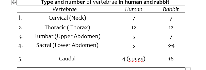
Cervical Vertebrae
- These are found in the neck region of a mammal.
- The distinguishing feature is a pair of verte-braterial canals in the neural arch, through which the blood vessels of the neck pass.
- Another feature is the structure of the transverse processes.
- They are flattened out and are known as cervical ribs.
- The fIrst cervical vertebra is known as the Atlas.
- It has a large neural canal and no centrum.
- The second cervical vertebra, is called axis.
- The other five cervical vertebrae have no specific names.
- They have the same structure.
- The cervical vertebrae possess numerous processes for muscle attachment.
Thoracic Vertebrae
- Each thoracic vertebra has a large centrum ,a large neural canal, neural arch and a long neural spine that projects upwards and backward.
- There is a pair of prezygapophyses and postzygapophyses for articulation with other vertebra .
- They have a pair of short transverse process.
- The thoracic vertebra also articulates with pair of ribs at tubercular and capitular facets.
Lumbar Vertebrae
- Each lumbar vertebra has a large, thick centrum for support of the body.
- It has a neural spine that projects upwards and forwards.
- There is a pair of large transverse process that are directed forwards.
- Above the prezygapophyses lies a pair of processes called metapophyses,
- Below postzygapophyses lies the anapophyses.
- Metapophyses and anapophysis serve for attachment pf muscles of the abdomen.
- In some mammals, there may be another process on lower side of centrum called hypapophysis also for muscle attachment.
Sacral Vertebrae
- The sacral vertebrae are fused together to form a rigid bony structure, the sacrum.
- The centrum of each vertebra is large, but the neural canal is narrow.
- The neural spine is reduced to a small notch.
- The transverse processes of the first sacral vertebra are large and wing-like
- They are firmly attached to the upper part of the pelvic girdle.
Caudal Vertebrae
- Human beings have only four of these vertebrae which are fused together to form coccyx.
- Animals with long tails have many caudal vertebrae.
- A typical caudal vertebra appears as a solid rectangular mass of bone.
- The entire bone consists of the centrum only.
Appendicular Skeleton
- The appendicular skeleton consist of the limbs and their girdles.
Bones of Fore-limbs
Pectoral girdle
- Pectoral girdle is made of scapula, coracoid and clavicle.
- A cavity known as glenoid cavity occurs at the apex of the scapula.
- The humerus of the fore limb fits into this cavity.
- The clavice is a curved bone connecting the scapular to the sternum.
Humerus
- Humerus is found in the upper arm.
- It articulates with the scapula at the glenoid cavity of the pectoral girdle and forms a ball and socket joint.
Ulna and radius
- These are two bones found in the forearm.
- The ulna has a projection called olecranon process and a sigmoid notch which articulates with the humerus.
Bones of hind limb
Pelvic Girdle
- The pelvic girdle consists of two halves fused at the pubic symphysis.
- Each half is made up of three fused bones:
- the ilium,
- ischium
- Each half has cup-shaped cavity for the acetabulum for articulation with the head of the femur.
- Between the ischium and pubis is an opening obturator foramen where spinal nerves, blood vessels and a tough inflexible connective tissues pass.
- The ilium, ischium and pubis are fused to form the innominate bone.
The Femur
- The femur is the long bone joining the pelvic girdle and the knee.
- The head of the femur articulates with acetabulum forming the ball and socket joint at the hip.
- The femur has a long shaft.
- At the distal end it has condyles that articulate with the tibia to form a hinge joint at the knee.
- The patella covers the knee joint and prevents the upward movement of the lower leg.
Tibia and Fibula
- The tibia is a large bone, and the fibula a smaller bone is fused to it on the distal part.
- In humans the tibia and fibula are clearly distinguishable.
Joints and Movement
- Ajoint is a connection between two or more bones.
- Joints provide articulation between bones making movement possible.
- However some joints do not allow any movement e.g. the joints, between bones of the skull.
- Movable joints are of three main types:
Gliding joint
- g., joints which occur between the vertebrae wrists and ankles.
- The ends of the bones that make the joint are covered with cartilage.
- The bones are held together by tough ligaments.
Synovial joint
- The joint is enclosed by fibrous capsule lined by synovial membrane which secretes synovial fluid into the synovial cavity.
- The synovial fluid lubricates the joint.
- They are called synovial joints.
- They include hinge joint and ball and socket joint.
Hinge joint
- g. knee joint.
- The joint allows movement in one plane.
Ball and socket joint.
- e.g., hip joint.
- The joint allows rotation in all directions.
Types, Locations and Function of Muscles
- There are three types of muscles, located at various parts of the body.
- In order to function all use energy in form of ATP.
- These include smooth, skeletal and cardiac muscles.
Smooth Muscle (Involuntary Muscles)
- These are spindle-shaped and contain filaments with myofibrils.
- Each muscle is bound by plasma membrane.
- They are found lining internal organs such as alimentary canal, bladder, and blood vessels.
- They are controlled by involuntary part of the nervous system.
- They are concerned with movement of materials along the organs and tubes.
- They contract slowly and fatigue slowly .
Skeletal Muscle (striated or voluntary muscle)
- Skeletal muscles are striated and have several nuclei.
- They are long fibres each containing myofibrils and many mitochondria.
- They have cross-striations or stripes.
- They are also called voluntary muscles because the contraction is controlled by voluntary nervous system.
- They are surrounded by connective tissue and are attached to bones by tendons.
- Their contraction brings about movement of bone, resulting in locomotion.
- They contract quickly and fatigue quickly.
Cardiac Muscle
- Consist of a network of striated muscle fibres connected by bridges.
- Are short cells with numerous mitochondria and uninucleate.
- They are found exclusively in the heart.
- Contractions of cardiac muscles are generated from within the muscles and are rhythmic and continuous hence they are myogenic.
- They do not tire or fatigue.
- The rate can be modified by involuntary nervous system.
- Their contractions result in the heart pumping blood.
Role of muscles in movement of the human arm
- Muscles that bring about movement are antagonistic, i.e. when one set contracts the other relaxes.
Antagonistic muscles of human forelimb
- The biceps muscles of the forelimb act as flexors while the triceps muscles act as extensors.
- The biceps has its point of origin on the scapula and the point of insertion on the radius.
- The triceps has its points of origin on the scapula and humerus and is inserted on the ulna.
- When the muscles contract, the limb acts as a lever with the pivot at the joint.
- Contraction of biceps muscles bends (flexes) the arm while contractions of triceps extends the arm.
Practical Activities
To observe prepared slides of transverse section of stems of herbaceous and woody plants.
- Permanent slides of transverse sections of:
- Herbaceous plant and Woody plant are obtained.
- The permanent slide of a herbaceous plant is placed onto the stage of the microscope.
- Observations under the low power and medium power objective is made.
- A plan diagram is drawn and labelled.
- The permanent slide of a woody plant is placed on the stage of the microscope.
- Observations under the low power and medium power objectives are made.
- A plan diagram is drawn and labelled.
- In both cases, support tissues such as parenchyma, collenchyma, sc1erenchyma and xylem are observed.
To observe wilting in young herbaceous plants.
- A herbaceous potted plant e.g. bean plant is obtained.
- The plant is placed on the bench near a window and left for 3 days without watering on the third and subsequent day.
- The shoot droops due to fall in turgor pressure; caused by water loss.
To examine the exoskeleton in an arthropod.
- Obtain a beetle and observe the external structure.
- The exoskeleton is on the outer surface with muscles attached on inner side.
- The exoskeleton is hardened by chitin.
- Movement is due to joints on the limbs.
- Also examine various shed cocoons of insects e.g., butterfly.
To observe the external features of a finned fish.
- Fresh Tilapia is obtained and placed on a tray.
- Observations are made on the external features of the fish.
- A labelled drawing is made.
- Features like scales, fins a streamlined body and an operculum are seen.
- Opened operculum reveals the gills.
To examine bones of the axial skeleton of a rabbit.
- Bones of the vertebra column are obtained.
- These are cervical, thoracic, lumbar and sacral.
- For each of the bones the distinguishing features are listed down.
- Labelled drawings of the anterior and lateral views is made.
To observe bones of appendicular skeleton.
- Bones of pectoral girdle and fore limb are obtained i.e., scapula, humerus, ulna and radius.
- Labelled drawing of each bone is made.
- Observations on how the bones articulate with one another is made.
- Bones of pelvic girdle and hind limbs are obtained i.e., pelvic girdle, femur, tibia and fibula.
- Labelled drawings of each, bone is made.
- The distinguishing features of each bone is noted.
- Observations on how the bones articulate with one another is made.