
PART I: EXCRETION
- Excretion is a process by which living organisms separate and eliminate waste products of metabolism from their bodies
- Metabolic wastes are toxic to cells if allowed to accumulate, and could also alter the normal conditions for cell function, killing the cells
- Secretion is the release of certain useful substances produced by cells, such as enzymes and hormones
- Egestion is the removal of indigestible and undigested materials from food vacuoles or alimentary canal
EXCRETION IN PLANTS
- Plants lack complex excretory organs because:
- Their wastes accumulate very slowly
- The main wastes originate from carbohydrate metabolism and are often reused by the plant e.g carbon (IV) oxide, oxygen and water
- Wastes in form of gases are removed by simple diffusion thro stomata and lenticels
- Some wastes are stored in non toxic forms in certain tissues or organs which later drop off with age e.g resins, alkaloids
Economic Importance of Plant Excretory Products
- Tannins in barks of Acacia are used in leather tanning
- Caffeine in coffee berries and tea leaves are used to prepare stimulant drinks
- Quinine from the bark of cinchona tree and Aloe leaves is an antimalarial agent
- Papain from raw pawpaw fruit skins is a proteolytic substance used in food industry to soften beef
- Colchicine is an alkaloid used to induce non-disjunction in genetic breeding expts and to treat cancer
- Rubber is made from latex of rubber plant, used in making tyres and shoes
Excretion and Homeostasis in Unicellular Organisms
- Protozoans like Amoeba, Paramecium, remove CO2 and nitrogenous wastes by simple diffusion across their bodies. This is possible because these organisms have large surface area to volume ratio, which allows diffusion rate to be very fast. Wastes diffuse along a diffusion gradient from the cytoplasm across the cell membranes into the surrounding water medium
- Protozoans remove excess water and dissolved solutes from their bodies by using their contractile vacuole. These substances accumulate in the contractile vacuole, which swells and moves towards the cell membrane then bursts releasing the contents to the outside
EXCRETION IN ANIMALS
- Animals need an elaborate system of excretion because they have more complex bodies and have numerous cells and thus diffusion alone is not enough to remove wastes
- Excretory tissues and organs include flame cells in flat worms, nephridia in round worms, malpighian tubules in insects, gills, lungs , liver, kidneys and skin in vertebrates
The Mammalian Skin
- The main functions of the skin are:
- Defence: protective barrier against entry of microorganisms, mechanical damage, and UV from the sun
- Regulation of body temperature
- Excretion: salts, excess water, some urea
- Reception of stimuli: heat, cold, pain, touch, pressure
- Vitamin D synthesis on exposure to sunlight
- Fat storage in adipose tissue
Structure-Function Relationship of the Mammalian Skin
- Cornified layer: outermost layer of the skin is made of flattened dead cells filled with tough flexible substance called keratin, protects against mechanical damage and entry of bacteria, and reduces loss of water by evaporation
- Granular layer is the middle layer of skin made of living cells with granules, it gives rise to the cornified layer
- Malpighian layer is the innermost layer of the skin, made up of actively dividing cells that give rise to new epidermis, the cells have pigment granules called melanin that gives colour to skin, and also absorbs UV rays that are harmful to skin
- Blood vessels supply nutrients and oxygen to skin tissues and remove waste products, blood also regulates body temperature
- Lymphatic vessels drain excess tissue fluid
- Nerve endings detect external stimuli, creating awareness within the body to initiate appropriate responses
- Sweat glands consist of coiled secretory cells extending into long tubules that open to the surface of the skin as sweat pores. The cells absorb excess water, mineral salts, some urea, lactic acid and CO2 from the surrounding cells and tissues, and secreted to form sweat. This flows through the sweat duct into the skin surface. Sweat glands are involved in temperature regulation thro loss of water by evaporation from skin surface
- Hair shafts originate from hair follicles located in the Malpighian layer. Erector pili muscles attached to the follicle at one end and to the epidermis on the other end contract and relax to alter the angle btn the hair shaft and the skin. This varies the amount of air trapped btn the hair and the skin, which is important in temperature regulation
- Sebaceous glands are closely associated with hair follicles; they secrete sebum to keep hair and epidermis flexible and waterproof. Sebum contains antiseptic substances to protect against bacteria.
- Subcutaneous layer is a layer of fat beneath the dermis; it binds the skin to muscles and other organs deep in the body, stores fats and insulates against heat loss.
The Mammalian Kidneys
Structure and Function of the Kidneys
- The functions of kidneys are excretion, osmoregulation, ionic balance and regulation of pH
- Mammals have 2 kidneys located at the dorsal part of the abdominal cavity in the lumbar region
- Above each kidney are the adrenal glands that secrete hormones
- A kidney is convex on one side and concave on the other side. The concave side has a depression called hilum where renal artery enters to supply blood and renal vein leaves to remove blood
- From the hilum also leaves a large thick walled tube called ureter that links the kidney to the urinary bladder. The ureter transports urine from the kidney to the bladder, which stores urine for a short time. When the bladder is full, sphincter muscles located at the base of the bladder relax and urine is released via the urethra
- In males the urethra is long and joined to the reproduction system unlike in females
- LS of kidney show cortex, medulla and pelvis starting from the convex side towards the concave side. In the cortex are glomeruli and Bowman’s capsules, while in the medulla are Loops of Henle and collecting tubules, and in the pelvis are collecting ducts.
- The basic functional unit of the kidney is called the nephron
Fine Structure of the Nephron
- Each nephron is made up of two main parts namely a renal tubule and glomerulus
- A renal tubule has 5 main parts: Bowman’s capsule, proximal convoluted tubule, Loop of Henle, distal convoluted tubule and collecting tubule
- The glomerulus is a fine network of blood capillaries enclosed by the Bowman’s capsule. It is formed from the afferent blood vessel, a branch from the renal artery
- The efferent arteriole collects blood from the glomerulus and extends to renal tubule where it divides into capillaries that form a meshwork over the entire tubule. This blood is then collected by the venules that join into the venous system
The Role of Nephron in Excretion
- Kidneys receive blood from the renal artery
- This blood is rich in nitrogenous wastes e.g urea; and contains dissolved food substances, plasma proteins, mineral ions, hormones and oxygen
- The afferent arteriole entering the glomerulus is wider than the efferent arteriole leaving it, this creates resistance to blood flow and very high pressure in the glomerulus
- Furthermore, the renal artery branches directly from the aorta whose blood flow is at high pressure, contributing to the high pressure within the glomerulus
- This high pressure forces the liquid part of blood and dissolved substances of low molecular sizes (e.g urea, glucose, salts, hormones, amino acids) to pass out of the glomerulus into the cavity of the Bowman’s capsule
- Large sized molecules in the plasma such as proteins and blood cells are not filtered out This process is known as ultrafiltration and the filtrate formed is called glomerular filtrate
- This filtrate flows into the proximal convoluted tubule (PCT)
- All glucose, amino acids, some water and mineral salts are actively reabsorbed against a concentration gradient. Active reabsorption requires energy.
- Some urea is reabsorbed to the blood down its concentration gradient until its concentrations in the filtrate and blood are equal, so in each pass through the kidneys half the urea is removed from the blood and half remains in the blood
- The filtrate then flows into the loop of Henle where salts especially sodium chloride are reabsorbed into the blood by active transport
- At the distal convoluted tubule, a controlled amount of water is reabsorbed into the blood by osmosis
- Water reabsorption is enhanced by the high osmotic pressure of the blood due to salt reabsorption at the loop of Henle, and secondly by a hormone known as antidiuretic hormone
- The filtrate in the collecting tubule becomes urine and flows down into the collecting duct. Urine flows into the urinary bladder through the ureter
- As urine accumulates, the urge to urinate is initiated and the sphincter muscles relax to pass out urine
NB: when the bladder is full, stretch receptors in the elastic walls send impulses to the medulla oblongata of the brain, which causes the sphincter muscles to relax, causing urination (or micturition). This is an involuntary reflex response that we can learn to control to a certain extent when we are young.
Large urine volume – stretch receptors in bladder A – Brain B – bladder sphincter
Stimulus Receptor Coordinator Effector
Urination
Response
A = Sensory neurone, B= Motor neurone
Describe how urine is formed in the mammalian kidney (14 marks)
Diabetes Insipidus
- Cause: pituitary gland not producing ADH or producing insufficient ADH
- Effect: kidney tubules unable to reabsorb water from GF; diuresis; dehydration of body; frequent thirst
- Treatment: replacing ADH/vasopressin, with a synthetic form of the natural hormone
Structure-Function Relationship of the Nephron
I: Glomerulus
- Afferent arteriole is wider than efferent arteriole to generate high pressure for the process of ultrafiltration
- Capillary walls have very small pores to prevent filtration of large sized molecules and to allow only filtration of small sized molecules
- Dense network of capillaries to increase surface area for ultrafiltration
II PCT
- Cells lining PCT have numerous mitochondria to generate energy for active transport
- Cells lining PCT have microvilli to increase surface area
- Long and highly coiled to increase area
- Highly coiled to reduce the speed of flow of filtrate to give more time for efficient reabsorption
- Well supplied with blood capillaries to deliver nutrients and oxygen
- One cell thick wall for fast movement of substances
III Loop of Henle
- U-shaped loop to create a counter-current flow between the filtrate and blood in vessels
- Descending limb is impermeable to ions but permeable to water to allow some water to move out by osmosis into the blood stream so as to make the glomerular filtrate more concentrated as it descends the loop
- Ascending limb is impermeable to water but permeable to ions and contains Na+ and Cl– ions pumps to actively transport these ions into the blood stream to make it highly hypertonic relative to glomerular filtrate
IV DCT
- Well supplied with blood capillaries to supply nutrients and oxygen
- One cell thick wall for fast movement of substances
- Cells lining DCT have numerous mitochondria to generate energy for active transport
- Cells lining DCT have microvilli to increase surface area
- Long and highly coiled to increase area for reabsorption
- Highly coiled to reduce the speed of flow of filtrate to give more time for efficient reabsorption
- numerous membrane pumps for active transport
The Liver
Functions of the liver
1. Deamination
- Excess amino acids are broken down to form ammonia, and the residue is fed into carbohydrate metabolism
- Ammonia is highly toxic; it is converted into the less toxic urea through a series of reactions called Ornithine cycle
- Urea is released into the blood stream and finally removed by kidneys
2. Detoxification
- Processes by which the liver convert toxic substances into harmless or less harmful substances
- Substances in drugs, foods or drinks are detoxified in the liver
- Enzyme catalase produced by all cells is broken down into harmless products i.e water and oxygen
3. Thermoregulation
- If body temperature tends to fall below norm, the hypothalamus stimulates the liver to increase its exothermic metabolic reactions to produce more heat
- If body temperature tends to rise above norm, the hypothalamus stimulates the liver to increase its endothermic metabolic reactions to consume more heat thereby lowering body temperature
4. Haemoglobin elimination
- Haemoglobin from worn out RBCs is broken down in the liver into urochrome and bilirubin
- Urochrome is eliminated by the kidneys, it gives urine a light yellow colour
- Bilirubin is excreted together with bile juice into the alimentary canal
5. Regulation of plasma proteins
- Plasma proteins namely albumin, fibrinogen, prothrombin and globulins/antibodies are mainly synthesised from amino acids in the liver
- Deamination and haemoglobin elimination also regulate the amounts of proteins
- Hb is broken down into haem and globin
- Globin is digested into amino acids
- Haem group is converted into biliverdin and bilirubin, transported to gall bladder and mix with bile juice. Later excreted through faeces giving it a brown colour
- Manufacture of red blood cells during foetal stage
- Formation and elimination of excess cholesterol
- Regulation of fat metabolism
- Storage of blood
- Storage of vitamins B,C,E,K and minerals
- Blood sugar regulation
PART II: HOMEOSTASIS
Definition: maintenance of a steady state of internal environment within the body, despite wide fluctuations in the external environment
- Internal environment: immediate surroundings of body cells, which is the tissue fluid
- External environment: immediate surroundings of the organism, which may be aquatic or terrestrial
- Internal envt factors that should be kept at steady state: osmotic pressure, pH, temperature, ionic level
Principles of Homeostasis
- Homeostasis works on a feedback mechanism: positive and negative
- Negative feedback: body responses that restore a condition which is in excess or in deficiency to norm
- homeostasis relies mainly on negative feedback mechanisms
- Positive feedback: body responses that lead to further deviation of a condition from norm either causing further deficiency or further excess
- Most positive feedback mechanisms are abnormal and result from a failure of negative feedback mechanisms. They often lead to disease states e.g diabetes
- Very few positive feedback mechanisms are beneficial e.g milk let down, uterine contractions during birth
- Norm or set point, a genetically predetermined level of a parameter that must be kept steady for proper functioning of cells e.g temperature, blood pressure, pH
- For an homeostatic mechanism to work normally, there must be receptors capable of detecting the change; a control mechanism capable of initiating the appropriate corrective measures; and effectors that can carry out these corrective measures
Q Explain the role of negative feedback in homeostasis in mammals. [4]
The Role of the Kidneys in Osmoregulation
- Osmoregulation refers to the maintenance of correct amounts of water and salts in the body, hence osmotic pressure
- Low osmotic pressure (OP) of tissue fluids will cause cells absorb water by osmosis, swell and possibly burst
- High OP will cause cells to lose water by osmosis and shrink
- Osmoregulation is mainly carried out by the kidneys and neuro-endocrine system ( the hypothalamus and pituitary gland)
1. Water Balance
- If OP rises due to dehydration: hypothalamus is stimulated and sends impulses to the (anterior) pituitary gland which releases more antidiuretic hormone, ADH, ( or vasopressin) into the blood. On reaching kidneys, ADH causes DCTs and CTs to become more permeable to water. Thus, more water is reabsorbed into the blood stream, thereby lowering OP and producing concentrated urine.
- If OP drops due to excessive rehydration, hypothalamus is stimulated and sends impulses to the (anterior) pituitary gland. The gland is less stimulated, thus less ADH is released into bloodstream. The kidney tubules become less permeable to water. Thus less water is reabsorbed into the blood stream and this raises the OP of blood and dilute urine is produced
2. Ionic Balance
- Ion levels (Na+, Cl–, K+, Mg2+, Ca2+, PO43-)must be regulated within narrow ranges for efficient functioning of body processes like protein synthesis, respiration, nervous co-ordination and muscle contraction
- When the level of sodium ions in the blood is low, adrenal glands produce more aldosterone into the blood. This hormone then stimulates the Loop of Henle and the gut to reabsorb more sodium ions into the blood, thereby raising their amount to norm. When sodium ions are reabsorbed into the blood, chloride ions follow in order to neutralise the charge on the sodium ions.
- When the level of sodium ions in the blood rises above optimum level, adrenal glands produce less aldosterone into the blood, which then stimulates the Loop of Henle and the gut to reabsorb fewer amounts of sodium ions into the blood, thereby raising their amount to norm
3. Regulation of pH
- Kidneys get rid of hydrogen ions by combining them with ammonia at the DCT to form ammonium ions. (Cells lining the DCT have an enzyme that enables them to produce ammonia from the amino acid glutamine when the conditions allow)
4. Regulation of Blood Sugar
- adrenal glands produce adrenaline during emergencies which is transported in the bloodstream to the liver where it stimulates the liver to increase the breakdown of glycogen into glucose
- This will increase available glucose for respiration, and thereby releasing more energy for the emergency
The Role of Liver in Homeostasis
- Blood Sugar Regulation
- If the sugar level in blood is above the norm or set-point i.e. 90mg per 100cm3 of blood, this stimulates the beta cells in islets of Langerhans of the pancreas to produce more insulin (glucose level is the stimulus)
- Insulin stimulates liver to convert glucose to glycogen and fat for storage;
- increase the oxidative breakdown of glucose into CO2, water and energy;
- inhibit the formation of glucose from glycogen/inhibit glycogenolysis;
- and to inhibit the formation of glucose from non-carbohydrate sources/ inhibit gluconeogenesis;
- Thus, glucose level is lowered back to the normal range
- On the other hand, when glucose level drops below norm, this stimulates the alpha cells in islets of Langerhans in pancreas to secrete more glucagon
- Which stimulates the liver to: increase glycogenolysis to form glucose;
- convert fats, proteins and amino acids into sugar (gluconeogenesis);
- and reduce oxidative breakdown of glucose;
- Thus, glucose level is raised to the normal range
- Adrenaline is produced by adrenal glands during emergencies
- It is transported in the bloodstream to the liver where it stimulates the liver to increase the breakdown of glycogen into glucose thereby releasing more energy for the emergency
- Deamination
- Excess amino acids are broken down to form ammonia, and the residue is fed into carbohydrate metabolism
- Ammonia is highly toxic; it is converted into the less toxic urea through a series of reactions called Ornithine cycle
- Urea is released into the blood stream and finally removed by kidneys
- Detoxification
- Processes by which the liver convert toxic substances into harmless or less harmful substances
- Substances in drugs, foods or drinks are detoxified in the liver
- Enzyme catalase produced by all cells is broken down into harmless products i.e water and oxygen
- Thermoregulation
- If body temperature tends to fall below norm, the hypothalamus stimulates the liver to increase its exothermic metabolic reactions to produce more heat
- If body temperature tends to rise above norm, the hypothalamus stimulates the liver to increase its endothermic metabolic reactions to consume more heat thereby lowering body temperature
- Haemoglobin elimination
- Haemoglobin from worn out RBCs is broken down in the liver into urochrome and bilirubin
- Urochrome is eliminated by the kidneys, it gives urine a light yellow colour
- Bilirubin and biliverdin are is excreted together with bile juice into the alimentary canal
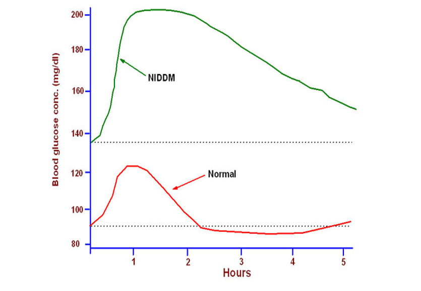
Diabetes Mellitus (DM)
- Diabetes mellitus is characterized by elevated blood glucose levels (hyperglycemia) and glucose in urine (glycosuria)
- It results from inadequate insulin secretion, or inability to respond to insulin, or both.
Type 1 Diabetes
- immune destruction of the beta cells of the pancreas
- Insulin secretion gradually diminishes
- May present at any age, but most common in childhood and adolescence
- Insulin by injection is necessary for survival
- Contributing factors: Genetic predisposition; environmental triggers (infection or other stress)
Type 2 Diabetes
- Caused by insulin resistance in the liver, increased glucose production in the liver, over production of free fatty acids by fat cells
- insulin secretion decreases with gradual beta cell failure
- Oral medication and/or insulin injections are eventually required.
- Contributing factors: Obesity, age (onset of puberty is associated with increased insulin resistance), lack of physical activity, genetic predisposition
Early Warning Signs for Type 1 and Type 2 Diabetes
- Increased urination
- Increased thirst
- Increased appetite
- Unexplained weight loss
Acid-Base Balance
- Blood and tissue fluids are kept at a pH of about 7.4
- This constancy depends on controlling the relative concentrations of acid and base
- This is achieved in 3 main ways:
- Lungs expel CO2
- Buffering mechanisms in the blood suppress the H+ ion concentration e.g. haemoglobin buffer, sodium hydrogen-phosphate buffer, sodium hydrogen carbonate buffer, plasma proteins buffer
- Kidneys get rid of hydrogen ions by combining them with ammonia at the DCT to form ammonium ions. (Cells lining the DCT have an enzyme that enables them to produce ammonia from the amino acid glutamine when the conditions allow)
Thermo-Regulation in Mammals
- Mammals are homoiotherms/endotherms: they maintain a constant body temperature despite wide fluctuations in temperature of external environment
- Internal body temperature is detected by a small region in the brain called hypothalamus as blood flows in the brain
- External environmental temperature is received by thermo-receptors in the skin, these relay the nerve impulse through sensory nerves to the hypothalamus
- The hypothalamus has numerous thermo-receptor cells to detect body temperature and initiate appropriate responses
- When body temperature tends to drop below norm:
- The hypothalamus sends impulses to the liver to increase exothermic reactions. More heat is produced and is distributed throughout the body by blood
- Blood vessels in the skin constrict (vasoconstriction occurs) to divert blood to a shunt system. This reduces blood flow to near the skin surface, which in turn decreases heat loss by radiation and convection
- Erector pili muscles are stimulated to contract, making hair to stand erect on the skin surface; trapping more air, which insulates the body against heat loss by radiation and convection
- Sweat glands reduce the rate of secretion of sweat, body heat is not used up to cause evaporation of water in sweat hence this conserves body heat
- In prolonged cold environmental temperatures, the skin stores more subcutaneous fat in the adipose tissue. Thick layer of fat offers more insulation against heat loss from the body
- Hypothalamus also initiates skeletal muscles start shivering which is involuntary contraction of muscles to generate heat during coldness
- Mammals also use behavioral activities to regulate body temperature during coldness.
- Some mammals hibernate i.e. go into deep sleep due to cold conditions. Others migrate to warmer habitats to avoid extreme cold weather
- Human beings put on heavy clothes and bask in the sun
- When body temperature rises above norm:
- Hypothalamus stimulates the blood vessels in the skin to dilate (vasodilation); increasing blood flow to near the skin surface; encouraging more heat loss by radiation and convection
- Sweat glands increase the rate of sweat production. Heat from the body is absorbed to cause evaporation of water in the sweat (latent heat of vaporisation) cooling the body
- Erector pili muscles relax making hair to lie flat on the skin surface; less air is trapped; reducing insulation; thereby encouraging more heat loss from the body surface by radiation and convection;
- Hypothalamus stimulates the liver to increase endothermic reactions; which consume body heat thus lowering body temperature
- Mammals living in persistently hot habitats have a thin layer of subcutaneous fat; reducing insulation; thus encourages more heat loss from the body
- Behavioural methods of lowering body temperature include migration to colder habitats, aestivation (going into deep sleep due to dry and hot conditions), moving into shade, and putting on light clothes
Thermoregulation in Plants
- Plants have the ability to tolerate wide changes in temperature
- Plants will increase transpiration rate when it is hot so as to cool down
- Shiny cuticle reflects heat
- A small leaf area reduces the uptake of heat
- Wilting during hot days reduces leaf surface area which is exposed to direct rays of the sun
NB: Wilting is caused by more water being transpired than can be replaced through the roots, resulting into the parenchyma cells losing turgidity, and the plant droops
Questions
- Describe the processes that prevent glucose being excreted in the urine
- Describe the role of insulin in the regulation of blood glucose concentration
- Describe how nitrogenous waste products are formed and explain why they need to be removed from the body.
- Describe how the kidney removes metabolic wastes from the body.
- Describe the part played by the proximal convoluted tubules in the functioning of the kidneys.
- Explain how the collecting ducts in the kidneys may reduce the loss of water from the body.